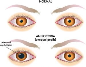

Anticholinergics such as atropine, homatropine, tropicamide, scopolamine, and cyclopentolate lead to mydriasis and cycloplegia by inhibiting parasympathetic M3 receptors of the pupillary sphincter and ciliary muscles. Pharmacologic anisocoria can present as mydriasis or miosis following administration of agents that act on the pupillary dilator or sphincter muscles. Causes include physical injury from ocular trauma or surgery, inflammatory conditions such as iritis or uveitis, angle closure glaucoma leading to iris occlusion of the trabecular meshwork, or intraocular tumors causing physical distortion of the iris. Mechanical anisocoria is an acquired defect that results from damage to the iris or its supporting structures. Examples include aniridia, coloboma, and ectopic pupil. Physiologic anisocoria may be intermittent, persistent, or even self-resolving.Ĭongenital anomalies in the structure of the iris may contribute to abnormal pupillary sizes and shapes that present in childhood. Light and near responses are intact, and the degree of anisocoria is typically equal in light and dark. The exact cause is unknown, but it is thought to be due to transient asymmetric supranuclear inhibition of the Edinger-Westphal nucleus that controls the pupillary sphincter. It is a benign condition with a difference in pupil size of less than or equal to 1 mm. Physiologic (also known as simple or essential) anisocoria is the most common cause of unequal pupil sizes, affecting up to 20% of the population. An injury or lesion in either pathway may result in changes in pupil size. Generally, anisocoria is caused by impaired dilation (a sympathetic response) or impaired constriction (a parasympathetic response) of pupils. Thus, thorough clinical evaluation is important for the appropriate diagnosis and management of the underlying cause. It is relatively common, and causes vary from benign physiologic anisocoria to potentially life-threatening emergencies. Anisocoria is a condition characterized by unequal pupil sizes.


 0 kommentar(er)
